Ushakaal Building, Nr Dhantoli Police Station

Dhantoli, Nagpur - 440012

10:30 AM - 9:00 PM

Monday to Saturday

Neuroimaging (Brain Sonography)
Neuroimaging-Brain-Sonography

What is Neurosonography?

It is an ultrasound technique by which high quality images of brain are obtained using an Ultrasonography machine with a specialized ultrasound probe / transducer. An ultrasound probe / transducer is placed on the head at specific locations, and returning signals are used to depict interior structures of brain.

It is an excellent modality for imaging brain in the infants and newborns. Neurosonography is also finding increasing use in other accessible regions of the central nervous system, including the adult brain during craniotomy and the spine during laminectomy.

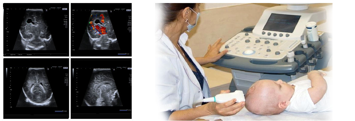
Clinical Uses of Neurosonography

  • Primary modality for the evaluation and screening of the brain in premature babies.
  • Allows detection and follow-up of brain complications which are usually seen in babies who have not cried immediately after birth (perinatal asphyxia) and premature babies.
  • Allows detection and follow-up of infants with hydrocephalus (fluid within brain ventricles).
  • Excellent anatomic imaging of the brain for evaluating for congenital anomalies in newborns and infants.
  • For evaluating children with brain infections / inflammatory processes such as ventriculitis, meningitis.
  • Neurosonography is also finding increasing use in the adult brain during craniotomy and the spine during laminectomy.

Advantages of Neurosonography (Brain Ultrasound) are

  • Safe, painless, non-invasive.
  • No ionizing radiation (in contrast to that encountered in x-ray and CT scan examinations), hence one of the safest of imaging techniques.
  • Quick.
  • Usually no sedation is required.
  • Allows real-time visualization of structures
  • Relatively inexpensive compared to other imaging modalities.