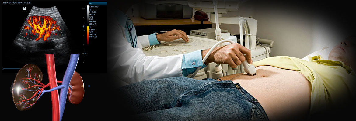
We, at Superspeciality Clinics & imaging Center offer you a wide range of genitor-urinary system Ultrasonography and Colour Doppler examinations at a fair cost and in a highly reliable & accurate manner.
Dhantoli, Nagpur - 440012
Monday to Saturday

Genito-urinary Imaging Services offered are:
- USG KUB (Kidney, ureter and Bladder)
- USG Prostate
- USG Pelvis
- Gynecological Imaging (USG of female pelvis including uterus and ovarian pathologies)
- TRUS (Transrectal Imaging): allows detailed imaging of Prostate & Seminal vesicles, also used for assessing vaginal vault recurrence in female patients treated for cervical cancer.
- TVS (Transvaginal Imaging) in female patients: Detailed assessment of uterus and ovaries and pelvic pathologies
- Ovulation study / Follicular Monitoring in patients with infertility
- USG scrotum: in patients with pain / swelling of scrotum, infertility, detection of undescended testis in children
A] USG KUB / renal ultrasounds when there's a concern about certain types of kidney or bladder problems. Renal ultrasound tests can show:
- The size of the kidneys
- Signs of injury to the kidneys
- Abnormalities present since birth
- The presence of blockages or kidney stones
- Complications of a urinary tract infection (UTI)
- Cysts or tumors
B] TRUS: A transrectal ultrasound of the prostate gland is performed to:
- Detect disorders within the prostate.
- Determine whether the prostate is enlarged, also known as benign prostatic hyperplasia (BPH), with measurements acquired as needed for any treatment planning.
- Detect an abnormal growth within the prostate.
- Help diagnose the cause of a man's infertility.
A transrectal ultrasound of the prostate gland is typically used to help diagnose symptoms such as:
- A nodule felt by a physician during a routine physical exam or prostate cancer screening exam.
- An elevated blood test result.
- Difficulty urinating.
Because ultrasound provides real-time images, it also can be used to guide procedures such as needle biopsies, in which a needle is used to sample cells (tissue) from an abnormal area in the prostate gland for later laboratory testing.
C] USG SCROTUM
Ultrasound imaging of the scrotum uses sound waves to produce pictures of a man’s testicles and surrounding tissues. It is the primary method used to help evaluate disorders of the testicles, epididymis (a tube immediately next to a testicle that collects sperm) and scrotum. Ultrasound is safe, noninvasive, and does not use ionizing radiation.
This procedure requires little to no special preparation.
What are some common uses of the procedure?
Ultrasound imaging of the scrotum is the primary imaging method used to evaluate disorders of the testicles, epididymis (a tube immediately next to a testis that collects sperm made by the testicle) and scrotum.
Common indications are:
- A mass in the scrotum felt by the patient or doctor
- Swelling of scrotum
- Trauma to the scrotal area.
- Causes of testicular pain or swelling such as inflammation or torsion.
- Evaluate the cause of infertility such as varicocele.
- Look for the location of undescended testis in neonates and infants.
A sudden onset of pain in the scrotum should be taken very seriously. The common causes of scrotal pain is epididymitis, an inflammation of the epididymes, which is treatable with antibiotics or torsion, commonly seen in adolescents, which if left untreated, can lead to loss of blood flow to the testicles and often requires surgical intervention.
D] USG PELVIS
What are some common uses of the procedure?
Ultrasound examinations can help diagnose symptoms experienced by women such as:
- Pelvic pain
- Abnormal vaginal bleeding
- Other menstrual problems
E] TRANSVAGINAL ULTRASOUND (TVS)
Allows more detailed evaluation of uterus, endometrium (the lining of the uterus) and the ovaries.
F] OVULATION STUDY / FOLLICULAR MONITORING:
It is a serial ultrasound examination usually by Transvaginal technique, which is used to monitor the formation and development of follicles within ovaries. It is done in patients with infertility.
